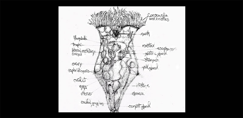
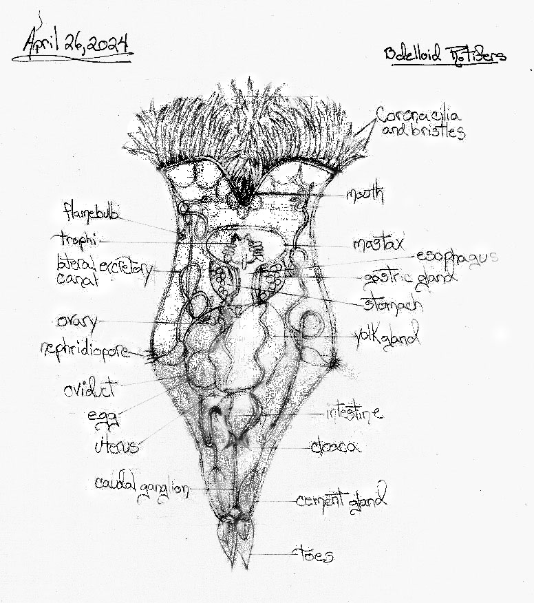
Flame Bulb:
A flame bulb, also known as a protonephridium, is a specialized excretory structure found in rotifers, including the Philodina Rotifer. It’s part of a larger system called the protonephridial system.
Each flame bulb consists of a cluster of cilia surrounded by a sac. The beating of the cilia creates a water current that draws waste products from the rotifer’s body cavity into the sac. From there, the waste products are transported through a network of tubules to the bladder and eventually expelled from the rotifer’s body.
Here are some key points about flame bulbs:
- They are not true kidneys, but they perform a similar function of osmoregulation and excretion.
- Each rotifer has one or two protonephridia, with each protonephridium containing multiple flame bulbs.
- The flame bulbs are located in the pseudocoelom, which is a fluid-filled cavity that surrounds the rotifer’s internal organs.
- The beating of the cilia in the flame bulbs is what gives them their name, as it creates a flickering appearance that resembles a flame.
Trophi:
In the context of rotifers, including the Philodina rotifer, trophi refers to their masticatory organs, also known as the mastax.
Trophi is plural, referring to the collective structure of several mouthparts working together. These mouthparts are responsible for grabbing food particles and grinding them up before they enter the rotifer’s digestive system.
Mastax:
A mastax is the muscular pharynx of a rotifer, like the Philodina rotifer, containing a complex chewing apparatus. It’s essentially a microscopic mouth with built-in jaws!
The mastax is a fascinating structure because its shape and composition vary depending on the rotifer species and its diet. Here’s a breakdown of its key features:
- Muscular walls: The mastax is surrounded by powerful muscles that allow it to crush and grind food particles.
- Trophi: These are the “jaws” of the mastax. They are made of hard, calcified materials and come in various shapes depending on the rotifer’s diet. For example, herbivorous rotifers may have trophi with grinding surfaces, while carnivorous rotifers may have trophi with sharp, grasping features.
- Function: The beating of cilia around the mouth opening creates a current that draws food particles into the mastax. The trophi then break down the food particles into a size suitable for digestion.
The mastax is a crucial part of the rotifer’s digestive system, as it allows them to efficiently consume a variety of food sources despite their microscopic size.
Lateral Excretory Canal:
In the context of a Philodina rotifer, there isn’t a structure directly called a “lateral excretory canal.” Philodina rotifers, like most rotifers, have a different excretory system compared to roundworms (nematodes) that possess lateral excretory canals.
Here’s the key difference:
- Roundworms (nematodes): They utilize a pair of lateral excretory canals as their primary excretory system.
- Rotifers (including Philodina): They rely on a protonephridial system for excretion. This system consists of flame bulbs (also called protonephridia) that filter waste products from the body cavity.
The flame bulbs have tubules that connect to a bladder, and waste products are expelled through a single opening. There aren’t paired canals running along the sides of the body like in roundworms.
Nephridiopore:
A nephridiopore is the external opening of a nephridium, which is an excretory organ found in many invertebrates, including rotifers like the Philodina rotifer. It essentially functions as a tiny drain for the organism’s waste products.
Here’s a breakdown of how it works:
- Nephridium: This is a tube-like structure that acts as the filtering unit of the excretory system.
- Filtration: Waste products and excess fluids from the body cavity pass through the nephridium’s walls.
- Nephridiopore: This is the opening at the end of the nephridium where the filtered waste products are expelled from the rotifer’s body.
Think of it like a miniature sewage system for the rotifer. The nephridium acts as the processing plant, filtering out waste, and the nephridiopore is the final exit point.
Here are some additional points to consider:
- The location of nephridiopores can vary depending on the specific invertebrate. In rotifers, the nephridiopore is usually located on the ventral side (belly) of the animal.
- Nephridia are similar to nephrons, which are the filtering units found in the kidneys of vertebrates like humans. However, nephridia are generally simpler in structure.
Yolk Gland:
Unfortunately, specific information about the presence and exact location of yolk glands in Philodina rotifers is limited in readily available scientific references.
Here’s what we know:
- Invertebrate reproduction: Philodina rotifers, like most invertebrates, likely rely on yolk for nourishing their developing embryos.
- Simpler reproductive system: Compared to vertebrates with dedicated yolk glands, Philodina rotifers likely have a more simplified approach to yolk production and storage.
Here are some possibilities:
- Internal yolk production: Yolk might be produced within the developing egg itself, eliminating the need for a separate yolk gland.
- Scattered yolk cells: There’s a possibility that yolk precursor substances are produced in scattered cells throughout the reproductive system and then incorporated into the developing eggs.
- Undocumented yolk gland: A less likely scenario is that Philodina rotifers do have a yolk gland, but its presence and specific location haven’t been extensively documented in general scientific references.
If you’re looking for very detailed information on yolk glands in Philodina specifically, you might need to consult more specialized research papers on the specific sub-species of Philodina rotifer you’re interested in.
Cloaca:
The cloaca in Philodina rotifers serves several important functions:
- Excretion: Waste products from the digestive system, as well as metabolic waste products, are expelled through the cloaca. This includes undigested food particles and other waste materials generated by cellular metabolism.
- Reproduction: In many species of rotifers, including Philodina, the cloaca also functions as the reproductive opening. During reproduction, eggs or embryos are released through the cloaca, and in some cases, sperm may also be deposited here during mating.
- Urinary System: While rotifers do not have true kidneys like more complex animals, they do have structures that help regulate osmotic balance and excrete excess water and salts. These waste products are also expelled through the cloaca.
- Gas Exchange: Although not its primary function, the cloaca may also play a role in gas exchange, allowing for the diffusion of gases such as oxygen and carbon dioxide across its membrane.
Overall, the cloaca in Philodina rotifers serves as a versatile and multifunctional structure, facilitating essential processes such as digestion, reproduction, excretion, and potentially even gas exchange, all within a single cavity.
Caudal Ganglion:
In the context of Philodina rotifers, the caudal ganglion refers to a cluster of nerve cells located at the posterior end of the animal, near the tail or caudal region. Ganglia are collections of nerve cell bodies that function somewhat like small brains, coordinating sensory information and controlling motor responses within specific regions of the body.
The caudal ganglion in Philodina rotifers plays a crucial role in coordinating movements and responses related to locomotion, particularly those involving the tail or posterior structures. It receives sensory input from the surrounding environment, such as touch or chemical cues, and integrates this information to generate appropriate motor responses. These motor responses may include movements of the tail or adjustments in body position to navigate the environment, avoid predators, or capture prey.
Overall, the caudal ganglion in Philodina rotifers is an important component of their nervous system, contributing to their ability to sense and respond to their surroundings effectively.
Cerebral Ganglion:
The cerebral ganglion, also commonly referred to as the brain of a Philodina rotifer, is the major nerve center responsible for coordinating its behavior and sensory perception. Here’s a breakdown of its key features:
- Location: It’s situated dorsally (towards the back) above the mastax (grinding mouthparts) and surrounded by either epithelial or muscular cells.
- Structure: The cerebral ganglion can vary in shape and size depending on the specific Philodina species, but it’s generally rounded, sac-shaped, or quadrangular.
- Function: It integrates sensory information from various parts of the rotifer’s body, such as eyes, antennae, and sensory bristles. Based on this information, the cerebral ganglion sends signals to muscles for coordinated movement, feeding, and escape responses.
- Neurons: The cerebral ganglion is packed with nerve cell bodies (perikarya) that extend nerve fibers (neurites) throughout the rotifer’s body. These neurites connect to muscles, sensory organs, and other tissues, forming a complex neural network.
Here’s some additional information to consider:
- Simple nervous system: Compared to vertebrates, the cerebral ganglion is a simpler structure. However, it allows rotifers to perform essential behaviors for survival and reproduction.
- Limited sensory organs: Philodina rotifers lack complex sensory organs but rely on simpler structures like photoreceptor eyes and touch-sensitive bristles to navigate their environment.
- Behavioral complexity: Despite the simple nervous system, Philodina rotifers can exhibit surprisingly complex behaviors, such as selective feeding, predator avoidance, and mating rituals. This highlights the efficiency of the cerebral ganglion in coordinating these actions.
Overall, the cerebral ganglion plays a vital role in the life of a Philodina rotifer, even though it’s a relatively simple structure compared to vertebrate brains. It allows them to interact with their environment, respond to stimuli, and carry out essential life functions.